Connectivity- vs Scalp-Based Targeting of Accelerated Transcranial Magnetic Stimulation for Depression: A Randomized Clinical Trial
Introduction
Major depressive disorder (MDD) remains a leading cause of disability worldwide, with many patients exhibiting resistance to standard treatments. Accelerated transcranial magnetic stimulation (aTMS) has emerged as a promising modality, offering rapid antidepressant effects through repeated sessions delivered over a short period. However, the optimal method for targeting brain regions during TMS remains under investigation. Traditionally, scalp-based landmarks such as the Beam F3 method guide coil placement over the left dorsolateral prefrontal cortex (DLPFC), a region implicated in mood regulation. Recent advances in neuroimaging—specifically functional connectivity mapping—offer the possibility to individualize targeting by identifying brain circuits most relevant to depression in each patient.
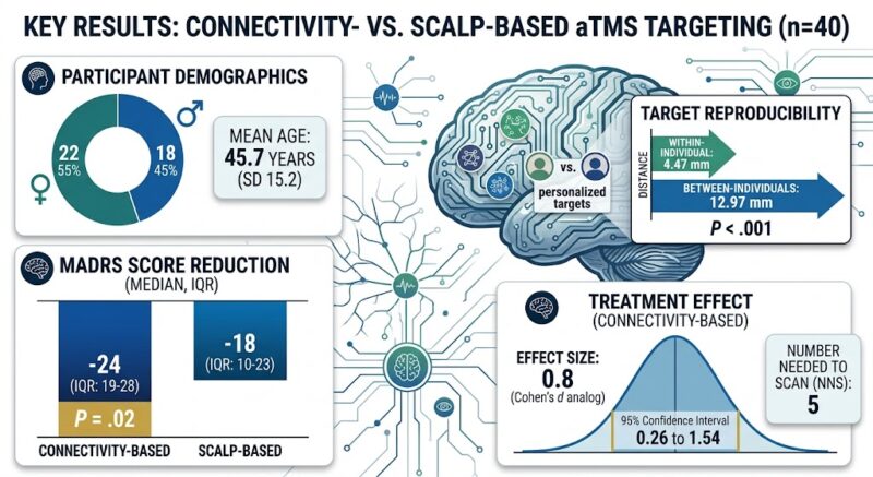
This study aimed to evaluate whether connectivity-based targeting of aTMS produces superior clinical outcomes compared to conventional scalp-based targeting in adults with treatment-resistant depression. Using resting-state functional MRI data, connectivity-based targets were individualized based on correlation with a well-characterized depression circuit involving negative connectivity to the subgenual cingulate cortex, a key node implicated in mood dysregulation.
Sign in free to continue reading
Create or use your MedXY account to unlock the complete article.
This article was created using several editorial tools, including AI, as part of the process. Human editors reviewed this content before publication.