Unraveling White Matter Hyperintensities: Distinct Lesion Subtypes Reveal Diverse Clinical Implications Beyond Location
Highlight
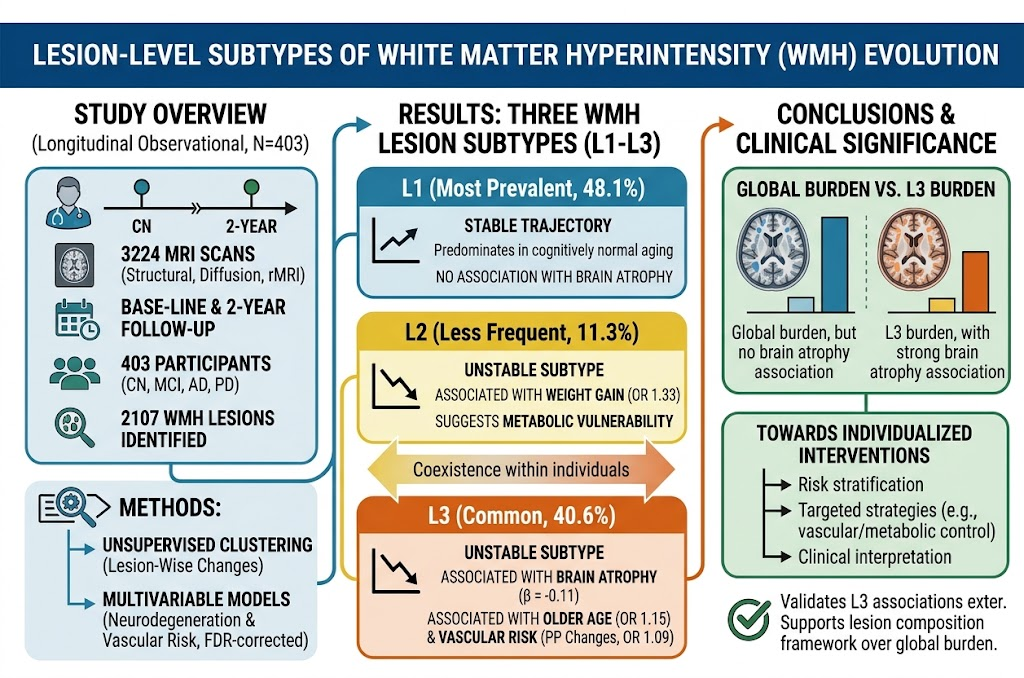
This longitudinal study identified three distinct lesion-level subtypes of white matter hyperintensities (WMHs) in elderly individuals with normal cognition, mild cognitive impairment, Alzheimer’s, and Parkinson’s disease. Each subtype demonstrated unique evolution patterns and associations: stable lesions unrelated to brain atrophy, unstable lesions linked to metabolic risk factors and weight gain, and unstable lesions associated with brain atrophy, older age, and vascular pulse pressure changes. Importantly, lesion subtype burden predicted neurodegeneration better than global WMH volume, underscoring biological heterogeneity beyond anatomical location.
Study Background
White matter hyperintensities (WMHs) are commonly observed on brain magnetic resonance imaging (MRI) in aging populations and patients with neurodegenerative diseases such as Alzheimer’s and Parkinson’s disease. Traditionally, WMHs have been quantified by total or regional burden, implicitly assuming homogeneous pathology. However, emerging evidence points to significant biological heterogeneity among individual WMH lesions, which may have distinct pathophysiological mechanisms and clinical relevance. Understanding this heterogeneity can clarify the cerebrovascular contributions to cognitive decline and brain atrophy and guide personalized risk stratification and therapeutic approaches.
Study Design
This large longitudinal observational study analyzed 3,224 MRI scans from 403 participants across a range of cognitive statuses—cognitively normal aging, mild cognitive impairment, Alzheimer’s disease, and Parkinson’s disease. MRI protocols included structural, diffusion-weighted, and resting-state sequences obtained at baseline and after 2 years. A total of 2,107 WMH lesions were identified and characterized longitudinally at the lesion level. Unsupervised clustering based on lesion evolution metrics was employed to define biologically meaningful WMH subtypes beyond anatomical location. Associations of subtype burden with neurodegeneration (quantified by brain atrophy measures) and vascular/metabolic risk factors were analyzed using robust multivariable regression models with false discovery rate (FDR) correction. Sensitivity analyses excluded anatomical location to confirm robustness, and findings were externally validated in an independent cohort.
Key Findings
Three distinct lesion subtypes were identified, frequently coexisting within individuals:
- L1 lesions: Constituted the largest proportion (48.1%) and predominated among cognitively normal participants. These lesions exhibited relatively stable volume and diffusion metrics over time and were not associated with brain atrophy, suggesting a relatively benign chronic state.
- L2 lesions: Accounted for 11.3% of lesions and represented an unstable subtype characterized by dynamic evolution. Notably, these lesions were associated with weight gain (odds ratio [OR] 1.33, 95% confidence interval [CI] 1.22–1.45; pFDR ≤ 0.001), implicating metabolic vulnerability in their pathophysiology.
- L3 lesions: Made up 40.6% of lesions and were unstable, showing progression linked to brain atrophy (β = –0.11, 95% CI –0.16 to –0.05; pFDR < 0.001). These lesions were associated with older age (OR 1.15, 95% CI 1.07–1.23; pFDR < 0.001) and vascular risk, specifically changes in pulse pressure (OR 1.09, 95% CI 1.03–1.15; pFDR = 0.006). Importantly, after accounting for L3 lesion burden, total WMH volume was no longer significantly linked to brain atrophy, highlighting the critical role of this subtype in neurodegeneration.
The presence of all three subtypes within individual brains underscores WMH heterogeneity. Sensitivity analyses excluding lesion location from clustering yielded consistent subtype classifications, and external validation replicated the association of the atrophy-related L3 subtype. These findings challenge the conventional view of WMHs as a uniform entity and emphasize the need for lesion-level characterization.
Expert Commentary
The elucidation of lesion-level WMH subtypes adds a valuable layer of granularity to cerebrovascular imaging biomarkers. This study’s integration of longitudinal multi-modal MRI and rigorous statistical modeling advances our mechanistic understanding of WMH evolution. Distinguishing stable, metabolically vulnerable, and neurodegeneration-associated WMH lesions offers potential for improved prognostication and targeted intervention strategies.
However, certain limitations warrant consideration. The study population, though sizeable and diverse in cognitive status, may not fully represent all demographic groups. The mechanistic underpinnings of each subtype remain to be elucidated by histopathologic and molecular studies. Additionally, clinical translation will require development of standardized imaging protocols and accessible algorithms for subtype detection in routine practice.
Future research should explore whether these lesion subtypes differentially respond to therapeutic interventions such as blood pressure control, lifestyle modification, or neuroprotective treatments. Integration with fluid biomarkers and genetic data could further refine subtype characterization and risk prediction.
Conclusion
This comprehensive longitudinal MRI study delineates three biologically distinct WMH lesion subtypes beyond spatial distribution, each with unique clinical and neurobiological correlates. Identifying lesion-level heterogeneity enhances understanding of the cerebrovascular contributions to brain aging and neurodegeneration and holds promise for personalized medicine approaches. Moving beyond aggregate WMH burden to lesion-specific profiling may better inform prognosis, clinical decision-making, and development of targeted therapies aimed at mitigating vascular-related cognitive decline and brain atrophy.
References
- Gonzalez-Gomez R, Tagliazuchi E, Campo CG, et al. Lesion-Level Subtypes of White Matter Hyperintensity Evolution Beyond Spatial Location. Neurology. 2026;107(6):e218472. PMID: 42659615.
- Wardlaw JM, Smith C, Dichgans M. Small vessel disease: mechanisms and clinical implications. Lancet Neurol. 2019;18(7):684-696.
- Debette S, Markus HS. The clinical importance of white matter hyperintensities on brain magnetic resonance imaging: systematic review and meta-analysis. BMJ. 2010;341:c3666.
- Prins ND, Scheltens P. White matter hyperintensities, cognitive impairment and dementia: an update. Nat Rev Neurol. 2015;11(3):157-165.
This article was created using several editorial tools, including AI, as part of the process. Human editors reviewed this content before publication.