Distinct Geographic Atrophy Phenotypes in AMD: Characterizing Growth Patterns and Clinical Implications
Highlight
- Identification of three distinct geographic atrophy (GA) phenotypes in age-related macular degeneration (AMD): unifocal foveal-involving, large coalescing multifocal, and small numerous multifocal.
- Quantitative analysis using artificial intelligence-based segmentation and Gompertz modeling reveals different growth rates and projected GA sizes among phenotypes.
- Presence of a peripapillary component associates with significantly faster maximum GA growth and larger ultimate GA lesion size.
- These phenotypic distinctions have clinical implications for prognostication and design/interpretation of AMD clinical trials.
Study Background
Age-related macular degeneration (AMD) remains a leading cause of irreversible vision loss globally, with geographic atrophy (GA), representing the advanced dry form, leading to progressive central vision decline. GA is characterized by degeneration of the retinal pigment epithelium and overlying photoreceptors, manifesting as well-demarcated areas of atrophy on fundus autofluorescence (FAF) imaging. Understanding variability in GA lesion morphology and progression kinetics is paramount for patient prognostication, personalized care, and effective clinical trial design. However, descriptions of GA phenotypes and their differential growth parameters remain limited. This study addresses this gap by characterizing candidate GA phenotypes and quantitatively analyzing their growth dynamics using longitudinal FAF data integrated with artificial intelligence (AI) segmentation and statistical modeling.
Study Design
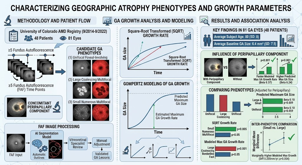
This prospective cohort study enrolled patients with GA secondary to AMD from the University of Colorado AMD Registry between September 2014 and September 2022, followed through April 2023. Inclusion required at least five longitudinal FAF imaging time points per eye to robustly assess lesion progression. Two independent graders reviewed each FAF image to classify GA lesions into one of three candidate phenotypes: (1) unifocal foveal-involving, (2) large coalescing multifocal, and (3) small numerous multifocal. Additionally, the presence of a peripapillary GA component was noted.
Each FAF image underwent automated GA lesion delineation using an AI-based segmentation model, followed by manual correction and validation by a vitreoretinal specialist to ensure accuracy. GA lesion area was measured, and a square-root transformation of lesion area was applied to stabilize variance for growth rate calculation. Gompertz modeling was employed to estimate each eye’s maximum GA growth rate and predict maximum future GA size over time. Linear regression with generalized estimating equations analyzed the association of GA phenotypes and peripapillary involvement with growth parameters.
Key Findings
A total of 81 eyes from 48 patients were analyzed, with a mean age of 80 years (SD 8). Baseline GA lesion area averaged 6.4 mm2 (SD 7.5), indicating diverse disease severity at study entry.
Peripapillary Component Impact: Eyes exhibiting a peripapillary GA component demonstrated significantly greater maximum modeled GA growth rates (Beta 0.37; 95% CI 0.16 to 0.59; p<0.001) and larger predicted maximum GA lesion size (Beta 0.24; 95% CI 0.10 to 0.38; p<0.001) in univariate analyses compared to those without such involvement. This highlights the peripapillary region as a site indicative of more aggressive GA progression.
Phenotypic Variations in Growth: After adjusting for peripapillary involvement, both large coalescing multifocal and small numerous multifocal phenotypes were associated with larger predicted maximum GA sizes compared to the unifocal foveal-involving phenotype (Beta 0.10; 95% CI 0.03 to 0.18; p=0.009 and Beta 0.10; 95% CI -0.00 to 0.21; p=0.059, respectively).
The small numerous multifocal phenotype had significantly faster square-root transformed (SQRT) and modeled maximum GA growth rates than the unifocal foveal-involving phenotype (Beta 0.09; 95% CI 0.03 to 0.16; p=0.005 and Beta 0.29; 95% CI 0.13 to 0.44; p<0.001). The large coalescing multifocal phenotype’s SQRT growth rate was borderline higher compared to the unifocal phenotype (p=0.05). Moreover, the small numerous multifocal phenotype showed a marginally higher modeled maximum GA growth rate than the large coalescing multifocal phenotype (estimated marginal mean difference 0.200 mm/year; 95% CI -0.00 to 0.40; p=0.054).
These results collectively indicate that multifocal GA phenotypes, especially the small numerous lesions subtype, may portend a more aggressive disease course with faster lesion expansion and potentially worse visual prognosis.
Expert Commentary
This study advances our understanding of GA heterogeneity in AMD by combining detailed phenotypic classification with robust longitudinal data analytics harnessing AI segmentation and Gompertz modeling, widely respected for modeling biological growth curves. Identification of distinct GA phenotypes with different growth kinetics enhances personalized prognostic counseling and may refine patient stratification for clinical trials testing emerging interventions.
The association between peripapillary atrophy and worse growth metrics aligns with hypotheses that GA contiguous to the optic nerve head may reflect broader retinal pigment epithelium dysfunction or vascular insufficiency. It remains essential to validate these phenotypic distinctions across diverse cohorts and imaging modalities. Future investigations incorporating genetic and environmental risk factors could elucidate mechanistic pathways influencing these growth patterns.
Study limitations include the single-center cohort, which may affect generalizability, and potential residual confounding despite adjustment for key variables. The requirement of multiple FAF time points restricts applicability to patients with frequent imaging follow-ups.
Conclusion
This prospective cohort study delineates three candidate geographic atrophy phenotypes in AMD, revealing significant differences in lesion growth rates and projected maximum lesion sizes. Multifocal phenotypes, particularly the small numerous multifocal type, and the presence of a peripapillary component are associated with more aggressive GA progression. These insights have significant clinical implications in improving prognostic accuracy and optimizing clinical trial design for emerging GA therapies. Continued research is warranted to validate these phenotypes and to explore underlying biological mechanisms driving differential growth trajectories in GA.
Funding and Clinical Trials
The study was supported by funding sources acknowledged in the original publication from the University of Colorado AMD Registry. No clinical trial registration was specified in the source publication.
References
1. de Carlo Forest TE, Mathias MT, Grove N, et al. Describing Candidate Geographic Atrophy Phenotypes and Their Different Growth Parameters. Am J Ophthalmol. 2026 Aug 20. PMID:42624312.
2. Sunness JS, Rubin GS, Applegate CA, et al. Reticular pseudodrusen and geographic atrophy in age-related macular degeneration. Retina. 2016;36(4):782-790.
3. Fleckenstein M, Charbel Issa P, Finger RP, et al. The progression of geographic atrophy secondary to age-related macular degeneration. Ophthalmology. 2018;125(3):369-390.
4. Schmitz-Valckenberg S, Fleckenstein M, Scholl HP, Holz FG. Fundus autofluorescence imaging in age-related macular degeneration. Baseline characteristics and lesions progression. Ophthalmology. 2009;116(10):1839-1846.
5. Holz FG, Sadda SR, Staurenghi G, et al. Imaging protocols in clinical studies in advanced age-related macular degeneration: Recommendations from Classification of Atrophy Consensus Meetings. Ophthalmology. 2017;124(4):464-478.
This article was created using several editorial tools, including AI, as part of the process. Human editors reviewed this content before publication.