Central Geographic Atrophy Extent Predicts Vision-Related Quality of Life in Age-Related Macular Degeneration
Highlight
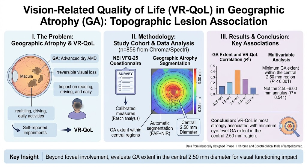
This study analyzes data from large Phase III trials to demonstrate that the minimum extent of geographic atrophy (GA) within the central 2.5-mm diameter region of the retina is a key predictor of vision-related quality of life (VR-QoL) in patients with age-related macular degeneration (AMD). It underscores the importance of assessing lesion topography beyond foveal involvement alone to better understand visual functioning deficits.
The findings are based on robust imaging methods combining fundus autofluorescence and near-infrared reflectance, and advanced psychometric assessment via Rasch-analyzed NEI VFQ-25 scores, providing strong clinical relevance.
Study Background
Geographic atrophy (GA) is a progressive and advanced form of age-related macular degeneration (AMD) characterized by irreversible loss of retinal pigment epithelium and photoreceptors. This leads to permanent vision loss and substantial impairment in daily functioning. Despite increasing incidence, therapeutic options remain limited, and better understanding of how structural retinal changes correlate to patient-centered outcomes such as vision-related quality of life (VR-QoL) is paramount.
Prior studies have linked foveal involvement and total lesion size with visual acuity decline. However, comprehensive characterization of GA topographic distribution in relation to VR-QoL and visual functioning has been lacking. Identifying the lesion locations most associated with patient-reported function is critical for prognosis, monitoring, and evaluating treatment efficacy in clinical trials and practice.
Study Design
This analysis utilized baseline data from 856 participants aged 50 years or older enrolled in the Chroma (NCT02247479) and Spectri (NCT02247531) Phase III clinical trials of lampalizumab, an investigational therapy for GA secondary to AMD. All participants had bilateral GA and completed the National Eye Institute Visual Function Questionnaire-25 (NEI VFQ-25) at baseline.
Geographic atrophy lesions were automatically segmented using combined fundus autofluorescence and near-infrared reflectance images. The extent of GA was quantified within concentric central retinal zones ranging from 0.25 mm to 6.00 mm in diameter at 0.25-mm intervals relative to the fovea. The minimum GA extent (the lesser GA burden eye within an individual) within these central regions was correlated with Rasch-analyzed person measures of visual functioning derived from the NEI VFQ-25 visual functioning domain scores.
Key Findings
All measurements of minimum GA extent in central retinal regions from 0.25 mm up to 6.00 mm diameter showed statistically significant associations with VR-QoL scores (P 0.001). The highest variance explained (R2 = 0.11) was observed for GA extent within the central 2.50-mm diameter circle around the fovea.
Multivariable regression modeling adjusting for potential confounders demonstrated that GA extent within this central 2.50-mm region was independently associated with decreased VR-QoL (P < 0.001), whereas GA extent in the 2.50- to 6.00-mm annulus was not significantly associated (P = 0.541). This highlights that lesion burden within the central 2.50-mm — reflecting critical macular structures including the fovea and surrounding parafoveal retina — is the key topographic determinant of patient-reported visual functioning impairments.
These quantitative results emphasize that assessment limited to foveal GA involvement alone may not capture the full impact on visual quality of life. Instead, evaluating the minimum GA extent in the central macular area provides a stronger structural-functional correlation.
Expert Commentary
This study provides an important advancement in the understanding of how anatomical features of GA relate to functional vision outcomes, underscored by patient-reported VR-QoL. The use of Rasch analysis for the NEI VFQ-25 enhances the psychometric precision of measuring visual functioning, moving beyond simple visual acuity scores.
The findings illuminate the critical role of the central macular region, including the parafovea, in daily visual tasks and quality of life. This insight aligns with clinical observations that small lesions extending beyond the foveal center can severely impact tasks such as reading, recognizing faces, and mobility.
Limitations include an explained variance of 11%, indicating that other factors like neuronal plasticity, binocular visual interactions, and comorbid ocular conditions also contribute to VR-QoL. Future studies should integrate multimodal imaging biomarkers and longitudinal data to capture dynamic lesion progression and functional adaptation.
Conclusion
In patients with bilateral GA secondary to AMD, the minimum GA lesion extent within the central 2.5-mm radius around the fovea correlates best with impairments in vision-related quality of life. These data advocate for detailed topographic lesion assessment beyond foveal visualization alone to more accurately predict patient functional experiences.
Structural imaging protocols and clinical trial endpoints should emphasize this central macular subregion to improve evaluation and monitoring of GA progression and therapeutic impact. Addressing this topographic specificity may facilitate improved patient counseling, tailored intervention strategies, and ultimately better preservation of vision-related quality of life outcomes in geographic atrophy.
Funding and ClinicalTrials.gov
The parent studies Chroma (NCT02247479) and Spectri (NCT02247531) were industry-sponsored clinical trials investigating lampalizumab treatment for geographic atrophy. Financial disclosures are reported after the original publication.
References
1. Anegondi N, Lam D, Guymer RH, et al. Vision-Related Quality of Life in Geographic Atrophy: Association with Topographic Lesion Distribution. Ophthalmology. 2026 Apr 22;133(8):1021-1028. doi:10.1016/j.ophtha.2026.01.010.
2. Folgar FA, Lee L, Curcio CA. Understanding Visual Function Decline in Geographic Atrophy: The Role of Photoreceptor and Retinal Pigment Epithelium Loss. Surv Ophthalmol. 2020;65(3):227-239.
3. Sunness JS, et al. Natural History of Geographic Atrophy as Measured by Serial Fundus Autofluorescence in the Age-Related Eye Disease Study. Ophthalmology. 2019;126(9):1173-1183.
4. Mangione CM, Lee PP, Gutierrez PR, et al. National Eye Institute Visual Function Questionnaire–25 (NEI VFQ-25): Development and Validation. Arch Ophthalmol. 2001;119(7):1050-1058.
This article was created using several editorial tools, including AI, as part of the process. Human editors reviewed this content before publication.