Introduction and Context
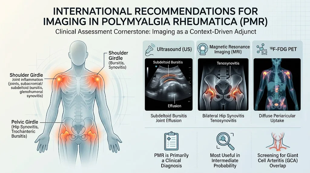
Polymyalgia rheumatica (PMR) is a common inflammatory disease of older adults characterized by bilateral pain and stiffness, typically involving the shoulder and pelvic girdles, and an often-elevated acute phase response. For decades clinicians have diagnosed and treated PMR largely on clinical grounds. However, advances in musculoskeletal ultrasound (US), magnetic resonance imaging (MRI), and 18F-fluorodeoxyglucose positron emission tomography (FDG-PET/CT) have offered tools to visualize the inflammatory features that can support or refute the diagnosis and sometimes reveal alternative causes of symptoms.
On October 27, 2026, an international, multidisciplinary task force published evidence-based recommendations for the use of imaging in the diagnosis and assessment of PMR (Bond et al., Ann Rheum Dis. 2026). The guidance—developed using the European Alliance of Associations for Rheumatology (EULAR) standard operating procedures—brings clarity about when imaging adds value and when it does not. These are the first international, consensus-based recommendations focused specifically on imaging in PMR and are intended for rheumatologists, radiologists, primary care physicians, and others involved in caring for older adults with new-onset proximal limb pain and stiffness.
Why these recommendations now
– Heterogeneous use of imaging across centers and specialties led to inconsistent diagnostic pathways and variable resource use.
– Rapid advances in US, MRI and PET technique and availability motivated an updated, evidence-based synthesis of their roles.
– Gaps existed in guidance for common clinical scenarios: atypical presentations, suspected overlap with large-vessel vasculitis (giant cell arteritis [GCA]), and whether serial imaging should guide therapy.
New Guideline Highlights
Major themes from the task force’s recommendations:
– PMR remains primarily a clinical diagnosis—imaging is an adjunct, not a replacement.
– Imaging is unnecessary when clinical pretest probability is clearly high or clearly low.
– Imaging is most useful when pretest probability is intermediate (atypical features, suspected alternative or overlapping conditions).
– No single imaging modality is endorsed as universally first-line; choice should be individualized by presentation, differential diagnoses, access, and local expertise.
– Vascular imaging (ultrasound, PET or cross-sectional angiography/imaging) is recommended when coexistence or overlap with GCA is suspected; routine screening for subclinical vasculitis is not supported.
– Using imaging routinely for disease monitoring or to predict outcomes is not recommended based on current evidence.
Key takeaways for clinicians
– Use clinical judgement as the primary diagnostic tool; reserve imaging for targeted questions (e.g., confirm uncertain diagnosis, exclude mimics, evaluate suspected large-vessel involvement).
– If imaging is performed, select modality according to the clinical question (see Topic-by-Topic Recommendations below).
– Do not order systematic vascular imaging in all PMR patients to look for subclinical GCA.
– Avoid routine repeat imaging to monitor response to therapy or to predict relapses—management decisions should be driven by clinical assessment and laboratory markers.
Updated Recommendations and Key Changes
What is new compared with prior practice and guidance?
– Formalized the concept of imaging only for intermediate pretest probability. Earlier practice was variable, with some clinicians imaging routinely and others infrequently.
– Explicit statement that no single modality is uniformly preferred as first-line. Prior literature often favored ultrasound for shoulder/hip evaluation; the panel emphasized local expertise and access instead of a universal hierarchy.
– Clear guidance on vascular imaging: perform targeted vascular imaging only when clinical suspicion for GCA exists (eg, new headache, jaw claudication, visual symptoms, constitutional symptoms, markedly asymmetric inflammatory markers, or imaging/clinical features suggestive of large-vessel involvement).
– A conservative stance on imaging for monitoring and outcome prediction—task force concluded current data are insufficient to recommend routine use for these purposes.
Evidence driving the updates
– Systematic literature review underpinning the recommendations found variability in sensitivity and specificity across modalities and across anatomical targets, and heterogeneous study designs and populations.
– No imaging modality achieved consistently high enough diagnostic accuracy to replace clinical judgment in all settings.
– Imaging adds most value when the clinical picture is unclear (intermediate probability) or when alternative diagnoses are actively suspected (eg, septic bursitis, crystal arthropathy, rotator cuff tear, or malignancy).
Topic-by-Topic Recommendations
Below are the task force’s principal recommendations, summarized for clinical use. These reflect consensus statements shaped by the literature review and iterative expert discussion.
Overarching Principles (summary)
– PMR is primarily a clinical diagnosis; consider imaging as an adjunct.
– The decision to image should be guided by clinical pretest probability and intended impact on management.
– Imaging decisions must account for local availability, expertise, cost and patient preferences.
Specific recommendations (7 key points)
1) Imaging is not required to confirm PMR when the clinical probability is high. If history, bilateral shoulder and pelvic girdle symptoms, morning stiffness, and raised acute phase markers strongly suggest PMR, start treatment and manage clinically without mandatory imaging.
2) Imaging is not necessary to exclude PMR in patients with low clinical probability. If presentation and labs do not support PMR, routine imaging to ‘look for PMR’ is not recommended.
3) Imaging is most helpful in intermediate pretest probability. When clinical features are atypical (eg, onset <50 yr, prominent peripheral synovitis, asymmetric symptoms), or when alternative or overlapping conditions are suspected, targeted imaging should be obtained to refine diagnosis.
4) No single imaging modality is universally first-line. Choose modality according to the clinical question:
– Musculoskeletal ultrasound: first-line when the question is whether classic periarticular inflammatory lesions exist (eg, bilateral subdeltoid/subacromial bursitis, biceps tenosynovitis, glenohumeral synovitis, hip synovitis or trochanteric bursitis). Advantages: bedside availability, low cost, no radiation, dynamic assessment. Limitations: operator dependent, limited for deep pelvic or axial disease.
– MRI: consider for deep structures, atypical axial symptoms, or when US is inconclusive and soft-tissue detail is required. MRI is sensitive for bursitis and joint synovitis but is less accessible and more costly.
– FDG-PET/CT: useful when there is concern for occult large-vessel vasculitis, systemic vasculitis, or alternative diagnoses such as malignancy; helpful to detect metabolic inflammation in vessels or in occult neoplasia. Limitations: radiation, cost, less availability, and variable sensitivity for smaller anatomic targets.
5) Vascular imaging should be performed when overlap with giant cell arteritis (GCA) is suspected. If patients have symptoms/signs suggesting cranial or large-vessel GCA (eg, new-onset headache, visual symptoms, jaw claudication, limb claudication, markedly asymmetric inflammatory markers, or clinical suspicion of LVV), targeted vascular imaging (temporal artery ultrasound, PET/CT, CT or MR angiography) is recommended.
6) Systematic screening for subclinical vasculitis in all PMR patients is not supported. Routine vascular imaging of asymptomatic PMR patients to detect occult large-vessel vasculitis is not recommended because evidence does not show clear benefit to patient outcomes and may lead to overdiagnosis and unnecessary interventions.
7) Imaging is currently not recommended for routine disease monitoring or for outcome prediction. Existing data do not support serial imaging to guide tapering or predict relapse, and imaging findings do not consistently correlate with clinical course or long-term outcomes.
Technical and operational considerations
– When using ultrasound, standardized scanning protocols and trained operators improve diagnostic yield. Assess bilateral shoulders (subdeltoid bursae, rotator cuff interval, biceps tendon sheath) and hips when symptoms suggest pelvic girdle involvement.
– When PET/CT is used to evaluate for large-vessel vasculitis or alternative diagnoses, interpret findings in the clinical context because FDG uptake can be seen in infection, atherosclerosis and malignancy.
– MRI protocols should include sequences optimized for soft-tissue contrast around shoulders and hips (eg, fluid-sensitive sequences) and dedicated vascular sequences where vasculitis is suspected.
Practical algorithms
– High clinical probability: treat and monitor clinically; consider imaging only if response to therapy is atypical.
– Low clinical probability: investigate alternate diagnosis without routine PMR-focused imaging.
– Intermediate probability / atypical features: perform targeted musculoskeletal US (if available); escalate to MRI or PET/CT if diagnostic uncertainty persists or to rule out mimics. If large-vessel symptoms are present or suspected, obtain vascular imaging.
Expert Commentary and Insights
What the task force emphasized
– “Imaging should serve a question” was a recurrent theme: tests should be ordered where results will change management.
– Training and standardization are critical, particularly for ultrasound, to reduce inter-operator variability and improve diagnostic performance.
– Resource stewardship is important—routine imaging of clear-cut cases leads to unnecessary costs and potentially confounding findings.
Controversies and areas of debate
– Some experts argued for wider use of ultrasound given its bedside convenience and growing evidence base; others highlighted operator dependence and variable access.
– The role of PET/CT remains debated: while powerful for detecting vascular inflammation and occult malignancy, PET/CT’s cost and limited availability restrict routine use.
– How best to define “intermediate pretest probability” in day-to-day practice remains a matter of clinical judgement; the task force did not prescribe a rigid scoring system.
Research priorities identified by the task force
– Prospective studies comparing imaging-guided diagnostic algorithms to clinical-only approaches, including health-economic analyses.
– Standardization of imaging definitions and scoring systems, especially for ultrasound in PMR.
– Studies on whether imaging-guided management (eg, using PET to detect LVV) improves long-term outcomes.
– Validation of imaging findings as predictors of relapse, steroid dependence, or complications.
Practical Implications
For clinicians
– Reinforce clinical assessment as primary; use imaging selectively to answer focused diagnostic questions that will influence treatment.
– If you lack local expertise in ultrasound or PET interpretation, rely on clinical judgment, and consider referral centers for difficult cases.
– When vascular involvement is suspected, arrange appropriate vascular imaging promptly because diagnosing GCA early can prevent serious complications such as vision loss.
For health systems and radiology departments
– Invest in training and standardization for musculoskeletal ultrasound if PMR imaging demand exists locally.
– Prioritize rapid access to vascular imaging for patients with suspected GCA.
– Avoid blanket policies that mandate imaging for all suspected PMR patients; instead, develop pathways that emphasize clinical triage and targeted imaging.
Patient perspective
– Patients should understand that imaging is not always needed and that the decision to image depends on how likely PMR is and what the imaging result would change about care.
– When imaging is done, clinicians should explain the choice of modality, its benefits, limitations, and possible incidental findings.
Application: Two brief clinical vignettes
Vignette 1 — Mary, 72
Mary has 3 weeks of sudden-onset bilateral shoulder and hip girdle stiffness worse in the morning, markedly elevated CRP, and rapid improvement after low-dose prednisone trial. Clinical probability of PMR is high. Recommendation: no routine imaging is required; treat and monitor clinically.
Vignette 2 — John, 65
John has unilateral shoulder pain, systemic weight loss, and only mildly raised inflammatory markers. Examination suggests possible rotator cuff disease, and there’s concern for occult malignancy given systemic features. Recommendation: targeted ultrasound of the shoulder (to evaluate bursitis, effusion, rotator cuff tear) and consideration of PET/CT if clinical and lab features raise concern for systemic disease or large-vessel inflammation.
References
– Bond M, Hysa E, Konzett V, et al. International recommendations for the use of imaging in the diagnosis and assessment of polymyalgia rheumatica. Annals of the Rheumatic Diseases. 2026 Oct 27. PMID: 42946585. https://pubmed.ncbi.nlm.nih.gov/42946585/
(Readers should consult the full task force publication for the detailed wording of the overarching principles, the seven recommendations, and the supporting evidence tables and voting results.)
Closing summary
These international recommendations represent a pragmatic, evidence-based recalibration of imaging practice in PMR. They reaffirm that clinical assessment remains the cornerstone of diagnosis, endorse imaging as a selectively applied adjunct in cases of diagnostic uncertainty or suspected overlap with GCA, and caution against routine use of imaging for monitoring or screening for subclinical vasculitis. Implementation will depend on clinician judgment, local resources and imaging expertise—but the overall message is clear: image to answer a question that matters for management, not as a reflexive diagnostic ritual.