Vessel Wall MRI in Pediatric Arterial Ischemic Stroke: Insights into Diagnosis and Prognosis
Highlight
- Vessel Wall MRI (VW-MRI) detects inflammatory changes in pediatric focal cerebral arteriopathy (FCA).
- Concentric vessel wall enhancement correlates with stenosis severity in FCA.
- VW-MRI shows limited prognostic value for clinical and radiological outcomes at 12 months.
- Enhancement likely reflects acute inflammation rather than long-term arterial pathology.
Study Background
Arterial ischemic stroke (AIS) in children is a significant cause of neurological morbidity with long-term consequences. Focal cerebral arteriopathy (FCA) is among the most common underlying arteriopathies in pediatric AIS and often involves inflammatory changes in the cerebral vessels. Accurately diagnosing the arteriopathy subtype is crucial for directing management and predicting outcomes. Vessel wall magnetic resonance imaging (VW-MRI) is an advanced imaging modality that visualizes vessel wall pathology such as inflammation and remodeling, providing information beyond luminal stenosis seen in conventional angiography. However, VW-MRI’s role in pediatric AIS, particularly its diagnostic accuracy and prognostic significance, remains underexplored. This gap has clinical implications because distinguishing active inflammation from chronic vessel changes may impact therapeutic decisions and prognostication.
Study Design
This retrospective ancillary study was conducted within the framework of the national KidClot cohort and included data from the Necker-Enfants Malades Hospital stroke database in Paris. The study population comprised 67 children diagnosed with arterial ischemic stroke who underwent VW-MRI within 3 months of stroke onset and had follow-up MRI at least 18 months later. Causes of stroke were classified using the Childhood Arterial Ischemic Stroke Standardized Classification and Diagnostic Evaluation (CASCADE). VW-MRI was used to evaluate intracranial arterial vessel wall enhancement (VWE). The study primarily focused on the subgroup with FCA, comparing VWE-positive and VWE-negative patients. Clinical variables, imaging metrics, and outcome measures including initial Pediatric National Institutes of Health Stroke Scale (PedNIHSS) scores and 12-month Pediatric Stroke Outcome Measure (PSOM) scores were analyzed. Statistical methods included chi-square and Fisher’s exact tests for categorical variables, Mann-Whitney U test for qualitative/ordinal variables, and Spearman’s correlation for continuous variables.
Key Findings
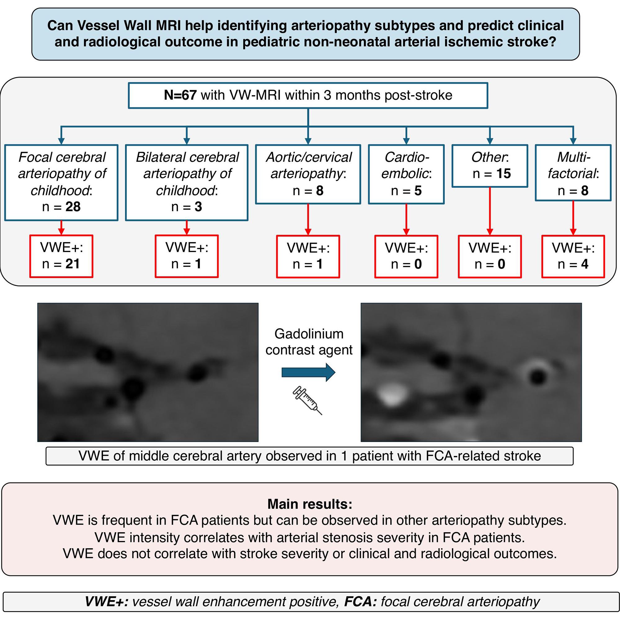
Among 67 children (median age 4.1 years; 61.1% male), FCA was the predominant cause, accounting for 42% (28/67) of cases. Other etiologies included bilateral cerebral arteriopathy (5%), aortic/cervical arteriopathy (12%), cardioembolic causes (7%), other (22%), and multifactorial causes (12%).
In the FCA subgroup, vessel wall enhancement was detected in 75% (21/28), predominantly concentric, a pattern suggestive of inflammatory involvement. The degree of VWE correlated positively with stenosis severity (Spearman ρ=0.31; P=0.001), indicating that more severe luminal narrowing is associated with greater wall inflammation. VWE-positive FCA patients had significantly higher initial arteriopathy severity scores (median 5 versus 3; P=0.046) relative to VWE-negative counterparts, suggesting that wall enhancement relates to disease burden of arterial narrowing.
However, VWE status was not significantly associated with initial stroke severity measured by PedNIHSS (median 8.5 versus 8; P=0.5) or functional outcome at 12 months assessed by PSOM (median 0.5 versus 1; P=0.8). This implies that while VW-MRI identifies active inflammatory vessel wall changes, these do not necessarily predict clinical severity at presentation nor long-term neurological deficits.
Expert Commentary
VW-MRI offers a detailed assessment of vessel wall pathology complementary to traditional vascular imaging. In pediatric AIS with FCA, detecting enhancement likely reflects acute inflammatory activity involving the intracranial vessels. This is biologically plausible given the inflammatory pathophysiology of FCA, often linked to post-infectious or immune-mediated processes.
Despite its diagnostic value, the limited prognostic impact observed in this study may be due to the complexity of stroke recovery influenced by multiple factors including collateral circulation, lesion size, and neural plasticity in children. Enhancement potentially readily resolves with treatment or natural course, thus not translating into long-term damage prediction. Additionally, limitations include the retrospective design, relatively small cohort, and the potential selection bias of patients undergoing VW-MRI.
Future directions should involve prospective, multicenter studies incorporating serial VW-MRI to characterize temporal changes in enhancement and its relationship to therapeutic interventions and neurological outcomes. Integration of VW-MRI findings with inflammatory biomarkers may also enhance prognostic capability.
Conclusion
VW-MRI is a valuable tool for detecting intracranial arterial wall inflammation in pediatric FCA post-arterial ischemic stroke, correlating with stenosis severity. However, it has limited ability to prognosticate clinical outcome at 12 months. Enhancement likely represents acute inflammatory changes rather than stable markers of long-term vessel damage or stroke prognosis. Thus, VW-MRI should be considered as a complementary diagnostic modality rather than a standalone prognostic indicator in pediatric stroke care.
Funding and Clinical Trials
The study was conducted using data from the national KidClot cohort. Specific funding sources were not detailed in the provided abstract.
References
1. Cazier P, Kossorotoff M, Naggara O, et al. Vessel Wall MRI in Pediatric Arterial Ischemic Stroke: Diagnostic Utility and Prognostic Value. Stroke. 2026 Sep 11; PMID: 42723611.
2. Amlie-Lefond C, Dlamini N, deVeber G. Arteriopathy in childhood arterial ischemic stroke: Results of the International Pediatric Stroke Study. Neurol Clin. 2010;28(3):585-602.
3. Gialdini G, Leary MC, Rosand J, et al. Arteriopathy diagnosis in childhood stroke: brain imaging and vessel wall imaging. Neurology. 2017;89(20):2049-2056.
This article was created using several editorial tools, including AI, as part of the process. Human editors reviewed this content before publication.