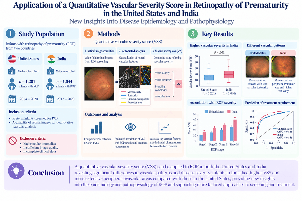
Highlight
– Application of an AI-derived vascular severity score (VSS) accurately correlates with ROP clinical staging and zone involvement.
– Comparative analysis of US and Indian cohorts reveals distinct vascular phenotypes despite similar clinical stages.
– Lower VSS values in Indian infants suggest geographic or pathophysiologic variations in ROP.
– Findings support the integration of quantitative imaging biomarkers with biochemical markers like VEGF for enhanced ROP understanding.
Study Background
Retinopathy of prematurity (ROP) is a vasoproliferative retinal disorder primarily affecting premature infants, characterized by abnormal retinal vascular development that can lead to visual impairment or blindness if untreated. Despite advances in neonatal care, ROP remains a significant cause of childhood blindness worldwide, particularly in low- and middle-income countries. The understanding of ROP epidemiology and pathophysiology varies geographically, influenced by differences in neonatal care, genetic predispositions, and screening practices.
Traditional ROP classification relies on clinical staging based on zone (location of disease), stage (severity of retinal changes), and extent of stage 3 (presence of extraretinal neovascularization). However, this qualitative approach may lack granularity and consistency across populations. Artificial intelligence (AI) technologies have recently enabled objective quantification of retinal vascular abnormality, offering promising tools for precision ROP assessment and monitoring.
Study Design
This retrospective comparative study evaluated two large data sets of ROP retinal images analyzed through the iROP deep learning (DL) system that outputs a vascular severity score (VSS) on a scale from 1 to 9, quantifying vascular abnormalities. The cohorts included:
A US multicenter cohort (iROP data set) encompassing 1019 infants and 6552 eye-level examinations collected between 2012 and 2020.
An Indian cohort from the Aravind Eye Care System (AECS), comprising 2363 infants and 8281 telemedicine retinal examinations from 2019 to 2020.
The study’s primary outcome was to explore associations between VSS and ROP clinical parameters, including zone, stage, and extent of stage 3 disease, using generalized estimating equations that accounted for repeated bilateral measures.
Key Findings
Analysis demonstrated that in both cohorts, a higher VSS consistently correlated with more severe ROP stages (from stage 1 to stage 3) and with increasing extent of stage 3 disease, confirming the score’s validity as an objective surrogate for vascular severity.
Moreover, for each ROP stage, greater posterior retinal zone involvement was linked with higher VSS, reflecting more severe posterior disease. These correlations held significance across almost all subgroup analyses in both populations.
Notably, when comparing cohorts at equivalent ROP stages, the AECS Indian cohort exhibited significantly lower mean VSS values than the US iROP cohort. This suggests phenotypic differences in ROP vascular manifestations beyond traditional clinical staging.
The authors hypothesize that these observed differences might stem from variable intraocular vascular endothelial growth factor (VEGF) levels influencing angiogenic activity and subsequent vascular severity measured by VSS. Although biochemical measures were not directly assessed, the vascular score may indirectly reflect underlying pathophysiologic mechanisms.
Parameter | US iROP Cohort | Indian AECS Cohort | Statistical Significance |
|---|---|---|---|
Number of infants | 1019 | 2363 | – |
Number of eye exams | 6552 | 8281 | – |
Mean VSS (stage 3) | Higher (mean score close to 9) | Lower | P < .001 |
Correlation of VSS with stage and zone | Strong positive correlation | Strong positive correlation | P < .001 |
Expert Commentary
These findings underscore the growing utility of AI-derived imaging biomarkers such as VSS in enhancing the objectivity and resolution of ROP disease assessment. The ability to quantify vascular severity complements traditional clinical staging, which can be subject to interobserver variability.
The distinct vascular severity patterns between the US and Indian populations highlight the complex interplay of environmental, genetic, and care-related factors influencing ROP pathophysiology. This has clinical relevance in tailoring screening protocols, intervention thresholds, and monitoring strategies adapted to local epidemiology.
The proposed relationship between VSS and intraocular VEGF aligns with current understanding that angiogenic factors modulate abnormal retinal neovascularization. Future integrated studies measuring both imaging biomarkers and molecular profiles could refine mechanistic insights and therapeutic targets.
Limitations include retrospective design, potential imaging quality variation, and absence of direct VEGF quantification. The generalizability to other regions remains to be validated.
Conclusion
The study robustly demonstrates that AI-derived vascular severity scoring effectively quantifies ROP severity correlating with established clinical parameters across diverse populations. Significant inter-population differences in vascular severity highlight heterogeneity in disease phenotype and pathophysiology, warranting deeper investigation.
Incorporating quantitative imaging markers like VSS into routine ROP evaluation promises to enhance diagnostic accuracy, enable personalized risk stratification, and improve outcomes globally. Integration with molecular biomarkers such as VEGF could further elucidate disease mechanisms and guide novel therapies.
Funding and Clinical Trial Registration
The original study was supported by relevant ophthalmology research foundations and institutions associated with the iROP and Aravind Eye Care System cohorts. Specific funding details and clinical trial registration numbers were not provided in the abstract.
References
Campbell JP, Nayak S, Ostmo S, et al. Application of a Quantitative Vascular Severity Score in Retinopathy of Prematurity in the United States and India: New Insights Into Disease Epidemiology and Pathophysiology. Am J Ophthalmol. 2026 Jun 13;290:123-136. doi:10.1016/j.ajo.2026.06.013. PMID: 42288327.
Gilbert C. Retinopathy of prematurity: a global perspective of the epidemiology and challenges in prevention and treatment. Early Hum Dev. 2008;84(2):77-82.
Campbell JP, Chiang MF. Advances in automated diagnosis of retinopathy of prematurity: pathophysiology insights and clinical applications. Curr Opin Ophthalmol. 2020;31(3):145-152.
Hartnett ME. Pathophysiology and mechanisms of severe retinopathy of prematurity. Ophthalmology. 2015;122(1):200-210.