Highlight
This prospective multicenter study demonstrates the high diagnostic accuracy of arterial spin labeling (ASL) MRI for detecting arteriovenous shunts (AVSs) in patients presenting with nontraumatic intracerebral hemorrhage (ICH). With a near-perfect negative predictive value, ASL reliably excludes AVSs such as arteriovenous malformations (AVMs) and dural arteriovenous fistulas (dAVFs), potentially serving as a noninvasive gatekeeper to digital subtraction angiography (DSA). The study reports excellent interobserver agreement and identifies ASL-positive shunts sometimes occult on initial DSA, highlighting its diagnostic strength and clinical utility in acute stroke etiologic workup.
Study Background
Intracerebral hemorrhage (ICH) represents a severe form of stroke characterized by bleeding within the brain parenchyma, carrying high morbidity and mortality. Secondary causes of spontaneous ICH include vascular malformations, predominantly arteriovenous shunts (AVSs) such as arteriovenous malformations (AVMs) and dural arteriovenous fistulas (dAVFs). Identifying these vascular anomalies is critical for tailoring patient management, guiding treatment interventions, and preventing rebleeding.
The diagnostic standard for AVSs remains digital subtraction angiography (DSA), an invasive procedure involving catheterization, ionizing radiation, and contrast administration, which may lead to complications and is not universally accessible. There is clinical need for a noninvasive, accurate imaging modality to screen for AVSs during the initial etiologic workup of acute ICH.
Arterial spin labeling (ASL) is a noninvasive MRI perfusion technique that uses magnetically labeled arterial blood water as an endogenous tracer, creating perfusion maps without radiation or contrast agents. Prior studies have suggested ASL can detect hyperperfusion associated with AVSs, but robust prospective data in unselected ICH populations are limited.
Study Design
This study was a prospective, multicenter diagnostic accuracy investigation conducted within the OPTICH cohort from October 2022 to October 2025 at three French tertiary hospitals. It enrolled consecutive adults with nontraumatic ICH who underwent standardized MRI, including ASL sequences, performed within seven days of hemorrhage onset.
Two experienced neuroradiologists independently and blindly assessed ASL perfusion maps, categorizing studies as positive for AVS if focal hyperperfusion adjacent to the hematoma was identified. The final ICH etiology incorporated all imaging modalities and follow-up, with mandatory reference standard DSA confirmation in all cases of AVS.
The primary endpoints were diagnostic performance metrics of ASL against the definitive etiologic diagnosis: sensitivity, specificity, positive predictive value (PPV), negative predictive value (NPV), and overall accuracy. Interobserver agreement was measured by Cohen’s kappa statistic.
Key Findings
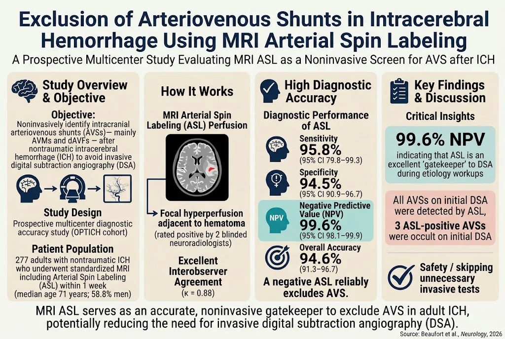
Out of 537 patients with nontraumatic ICH screened, 277 met inclusion criteria (median age 71 years; 58.8% male). Among these, 24 (8.7%) had confirmed AVSs (23 AVMs and 1 dAVF).
Interobserver agreement for ASL readings was excellent (κ = 0.88), attesting to the technique’s reproducibility. ASL achieved a sensitivity of 95.8% (95% CI 79.8–99.3) and specificity of 94.5% (90.9–96.7). The positive predictive value was moderate at 62.2% (46.1–75.9), while the negative predictive value was remarkably high at 99.6% (98.1–99.9). Overall diagnostic accuracy reached 94.6% (91.3–96.7).
Importantly, all AVSs identified on initial DSA were detected by ASL. Moreover, ASL revealed three AVS cases that were not apparent on initial DSA, underscoring its potential added sensitivity or detection of lesions missed by angiography at presentation.
The high NPV supports the use of ASL as a rule-out test: a negative ASL scan virtually excludes the presence of AVSs in an acute ICH context, potentially obviating unnecessary invasive DSA and reducing procedural risks and healthcare costs.
Expert Commentary
This study offers compelling evidence that arterial spin labeling MRI, a widely available and noninvasive method, can effectively exclude arteriovenous shunts in patients with spontaneous intracerebral hemorrhage. The prospective multicenter design and rigorous doped imaging adjudication lend credibility to the findings.
However, limitations exist. The selective use of DSA, primarily in patients with ASL-positive or clinically suspicious cases, may introduce a verification bias. The relatively small number of non-AVM shunts (only one dAVF) limits generalizability to all AVSs. Scanner heterogeneity, while potentially affecting image quality, reflects real-world practice and thus improves external validity.
These findings align with prior smaller-scale or retrospective studies that highlighted ASL’s sensitivity to focal hyperperfusion from AVMs or dAVFs. Mechanistically, AVSs create high-flow arteriovenous connections that manifest as localized hyperperfusion signals on ASL, providing a physiological basis for detection.
Current clinical guidelines recommend DSA as the gold standard for AVS diagnosis but emphasize the risks and resource demands associated with this modality. Incorporating ASL into routine MRI protocols may streamline the diagnostic pathway, reserving invasive angiography for positive or equivocal cases.
Conclusion
This prospective multicenter study demonstrates that arterial spin labeling MRI offers excellent diagnostic accuracy and a near-perfect negative predictive value to exclude arteriovenous shunts in patients with nontraumatic intracerebral hemorrhage. ASL’s noninvasive nature, coupled with its high reproducibility and ability to detect lesions sometimes missed by DSA, supports its integration as a gatekeeper in the acute ICH diagnostic workflow. Future external validation and cost-effectiveness analyses should further define the role of ASL in practice, potentially reducing reliance on invasive angiography, minimizing risks, and optimizing patient outcomes.
Funding and ClinicalTrials.gov
The study was conducted under the OPTICH collaborative framework, supported by institutional resources of the participating French tertiary hospitals. Specific funding details were not disclosed. The study is registered prospectively from October 2022 to October 2025; clinical trial registration information was not provided in the source text.
References
Beaufort Q, Agripnidis T, Amar V, Pandaure B, Bretonniere A, Cottier JP, L’Allinec V, Godard S, Codron P, Ifergan H, Cohen C, Pasi M, Boulouis G, OPTICH collaborators. Exclusion of Arteriovenous Shunts in Intracerebral Hemorrhage Using MRI Arterial Spin Labeling: A Prospective Multicenter Study. Neurology. 2026-10-29;107(10):e218705. PMID: 42956389. Available at: https://pubmed.ncbi.nlm.nih.gov/42956389/