Introduction
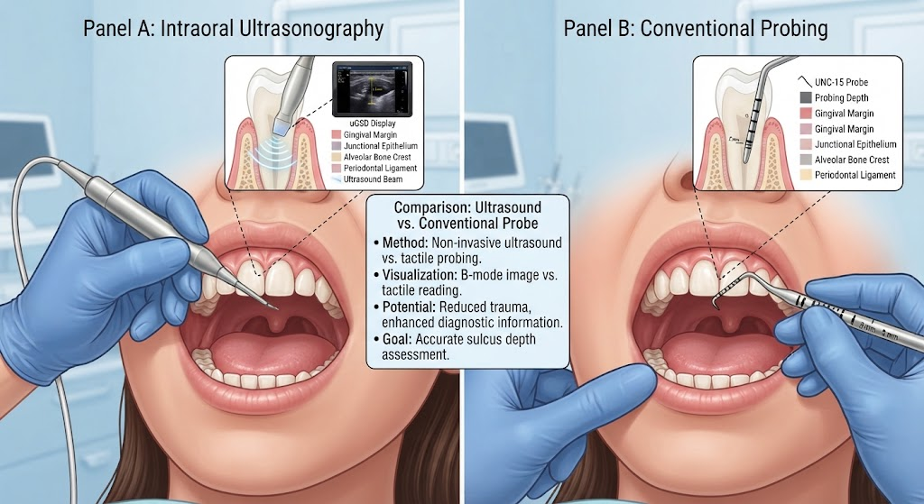
Periodontal health assessment is an essential component of diagnosing and managing gum diseases such as gingivitis and periodontitis. Traditionally, clinical probing depth (PD) measurements using manual probes like the UNC-15 are considered the gold standard for evaluating the depth of the gingival sulcus or periodontal pocket. These measurements inform treatment planning and disease monitoring. However, clinical probing is invasive and can cause patient discomfort, variability in measurements due to examiner technique, and potential tissue trauma.
In recent years, technological advances in ultrasonography offer a non-invasive alternative to traditional probing. Intraoral ultrasonography enables visualization of soft tissue structures through high-frequency sound waves, thus potentially estimating sulcus depth without tissue penetration. This study aimed to evaluate the agreement between ultrasound-derived gingival sulcus depth (uGSD) and clinical probing depth (PD) at standardized mid-buccal sites in anterior teeth, premolars, and first molars. The study’s goal was to establish feasibility and accuracy of intraoral ultrasound as a diagnostic adjunct.
Materials and Methods
The clinical agreement study enrolled 69 participants, encompassing a total of 963 measurement sites: 767 anterior teeth, 127 premolars, and 69 first molars. An 18-MHz intraoral ultrasound transducer was employed to capture high-resolution B-mode images of the gingival sulcus at the mid-buccal sites of these teeth. The ultrasound data was analyzed to measure the gingival sulcus depth (uGSD).
For comparison and validation, conventional clinical probing depth (PD) measurements were performed at identical sites using the standard UNC-15 periodontal probe. Two trained examiners independently measured uGSD to assess inter-examiner reliability.
Statistical analyses included intra-class correlation coefficient (ICC) assessments to evaluate reliability and agreement between uGSD and clinical PD. Bland-Altman plots assessed mean differences and limits of agreement. Mean absolute error (MAE) was calculated to quantify measurement discrepancies. Generalized estimating equations factored in clustering effects within patients. Subgroup analyses explored the influence of factors such as PD strata (shallow vs deep pockets), presence of bleeding on probing, tooth type, presence of supra- or subgingival calculus, restorations (prosthetic crowns or orthodontic attachments), and smoking status on measurement agreement.
Results
The study demonstrated excellent reliability for ultrasound measurements, with ICC values exceeding 0.90, indicating high reproducibility between examiners. Agreement between uGSD and clinical PD was also strong, with an overall ICC of 0.942 (95% confidence interval: 0.935-0.949).
The mean difference between ultrasound and clinical probing was minimal (+0.023 mm), reflecting negligible systematic bias. The mean absolute error (MAE) was low at 0.29 ± 0.32 mm, indicating close approximation between methods.
Subgroup ICCs ranged from 0.786 to 0.952, showing robust agreement across different clinical conditions and tooth types. Notably, 85.0% of measurement sites differed by 0.50 mm or less, and 96.7% differed by 1.00 mm or less, underscoring strong concordance.
Among tooth types, molar sites exhibited the highest MAE (0.38 mm) and a slight underestimation bias of -0.123 mm by ultrasound compared to probes. This may relate to anatomical complexities or imaging limitations in molar regions.
Discussion
This proof-of-concept study confirms that intraoral ultrasonography can reliably measure gingival sulcus depth with high agreement to conventional clinical probing at standardized mid-buccal sites. The non-invasive nature of ultrasound offers several advantages: eliminating patient discomfort associated with probing, reducing risk of tissue trauma and bleeding, and potential for operator-independent measurements once trained.
The demonstrated high reliability between examiners suggests that ultrasound could provide consistent diagnostic data, potentially reducing inter-examiner variability that challenges traditional probing methods. Furthermore, ultrasound imaging captures soft tissue morphology visually, providing richer diagnostic information than a simple numeric depth.
Limitations include slightly lower accuracy in molar areas, which may necessitate technical improvements in transducer design or imaging protocols. Moreover, the current study was limited to accessible mid-buccal sites in anterior teeth, premolars, and first molars. Comprehensive assessment across more anatomically complex posterior areas and interproximal sites would be valuable.
Examiner expertise also influences ultrasonography performance; widespread clinical adoption would require robust training programs. Additionally, cost and accessibility of intraoral ultrasound equipment versus handheld probes must be considered.
Future research should explore integration of this technique in routine periodontal diagnosis and monitoring, possibly combined with digital records and automated image analysis for enhanced clinical workflows.
Conclusion
Intraoral ultrasonography demonstrates high agreement and reliability in measuring mid-buccal gingival sulcus depth compared to standard clinical probing, with minimal measurement bias. This promising non-invasive modality could serve as an adjunct tool for periodontal assessment, improving patient comfort and measurement consistency.
Broader clinical studies are warranted to validate these findings across diverse populations, various tooth sites including complex posterior areas, and among clinicians with varying experience. Such research will clarify the full clinical utility and adoption potential of intraoral ultrasound in everyday dental practice.
Clinical Implications
Dentists and hygienists may soon have a valuable, patient-friendly alternative to manual probing that reduces discomfort and risk while providing accurate periodontal measurements. Adoption of intraoral ultrasonography could enhance early detection and monitoring of periodontal disease progression, facilitating timely interventions and improved oral health outcomes.
References
Aydindogan S, Ozdemir M, Balaban YE, Velioğlu EM, Hakki SS, Orhan K. Intraoral Ultrasonography for Mid-Buccal Probing Depth Assessment: A Proof-of-Concept Clinical Agreement Study. Journal of Clinical Periodontology. 2026 Oct 16. PMID: 42885429. Available at: https://pubmed.ncbi.nlm.nih.gov/42885429/