Highlight
Lower 24-hour systemic blood pressure, particularly minimum mean arterial pressure (MAP), systolic blood pressure (SBP), and diastolic blood pressure (DBP), is linked to faster retinal nerve fiber layer (RNFL) thinning in glaucoma.
24-hour ambulatory blood pressure monitoring provides valuable hemodynamic data for identifying glaucoma patients at higher risk of progression beyond traditional intraocular pressure assessment.
The study demonstrates a quantifiable association where every 10 mm Hg reduction in minimum 24-hour MAP results in an additional loss of approximately 0.54 µm per year of RNFL thickness.
Study Background
Primary open-angle glaucoma (POAG) is a leading cause of irreversible blindness worldwide. Its pathogenesis is multifactorial, with elevated intraocular pressure (IOP) being the primary modifiable risk factor. However, vascular factors, including systemic blood pressure (BP), have been implicated in disease progression. Low systemic BP, especially nocturnal hypotension or pronounced dips in BP, may compromise optic nerve head perfusion, potentially accelerating retinal ganglion cell loss. Despite this recognized association, longitudinal prospective data linking 24-hour BP metrics with objective markers of glaucomatous progression, such as retinal nerve fiber layer (RNFL) thinning measured by spectral-domain optical coherence tomography (SD-OCT), remain limited. The Vascular Imaging in Glaucoma Study aimed to clarify the relationship between ambulatory BP fluctuations and RNFL progression rates in patients with POAG.
Study Design
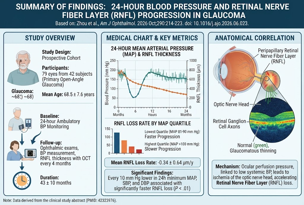
This was a prospective cohort study conducted at the Bascom Palmer Eye Institute. The analysis included 79 eyes from 42 subjects diagnosed with POAG, with a mean age of 68.5 years. Baseline 24-hour ambulatory BP monitoring was performed, capturing mean arterial pressure (MAP), systolic BP (SBP), diastolic BP (DBP), and their minimum values throughout the day and night. Ophthalmic follow-up evaluations were conducted every four months over an average of 43 months, including assessment of IOP, central corneal thickness, and RNFL thickness via SD-OCT. The primary endpoint was the rate of average peripapillary RNFL loss over time.
Linear mixed-effects models adjusted for confounding variables—age, sex, race, baseline RNFL thickness, central corneal thickness, and IOP—were used to assess the association between baseline ambulatory BP parameters and RNFL progression rates.
Key Findings
Over a mean follow-up period of approximately 3.5 years, multiple OCT scans per eye (average 13 ± 3) allowed robust longitudinal assessment of RNFL thinning. The mean RNFL loss rate was -0.34 ± 0.64 micrometers per year. Critical findings include:
For every 10 mm Hg lower minimum 24-hour MAP, there was a statistically significant increase in RNFL thinning rate by -0.542 µm/year (P < .001).
Likewise, reductions of 10 mm Hg in minimum SBP and DBP were associated with -0.360 µm/year (P = .003) and -0.458 µm/year (P = .008) faster RNFL progression, respectively.
Eyes in the lowest quartile of average 24-hour MAP (81-90 mm Hg) and minimum 24-hour DBP (35-47 mm Hg) showed significantly accelerated RNFL loss compared to those in the highest quartiles, with differences of -0.68 µm/year (P = .017) and -0.63 µm/year (P = .030), respectively.
These effects persisted after adjusting for known glaucoma risk factors, underscoring the independent influence of systemic BP on optic nerve head health.
Expert Commentary
The evidence from this study corroborates the pathophysiological hypothesis that ocular perfusion pressure, influenced by systemic BP variations, plays a critical role in glaucoma progression. The retinal nerve fiber layer serves as a sensitive structural marker for neurodegeneration in glaucoma, and its progressive thinning reflects cumulative ganglion cell loss.
Lower systemic BP, especially pronounced dips during the night, may reduce ocular perfusion pressure below the autoregulatory capacity of the optic nerve head vasculature. This could exacerbate ischemic injury and oxidative stress, precipitating faster structural damage. These findings highlight a limitation in current glaucoma management focusing largely on IOP control, suggesting the need for comprehensive cardiovascular assessment.
However, potential confounding factors such as antihypertensive medication effects, coexisting systemic vascular conditions, or nocturnal BP dipping patterns were not fully explored and warrant further study. Additionally, generalizability beyond the studied population requires confirmation.
Clinicians should consider 24-hour ambulatory BP monitoring as a valuable adjunctive diagnostic tool for risk stratification in POAG patients, particularly those progressing despite target IOP control.
Conclusion
In conclusion, the Vascular Imaging in Glaucoma Study provides compelling longitudinal evidence linking lower systemic blood pressure—most notably minimum 24-hour MAP, SBP, and DBP—to accelerated retinal nerve fiber layer loss in primary open-angle glaucoma. Integrating 24-hour ambulatory BP monitoring may enhance identification of individuals at elevated risk for disease progression, enabling tailored therapeutic strategies that address both ocular and systemic vascular factors.
Future research should aim to elucidate the mechanistic pathways connecting systemic hypotension and glaucomatous optic neuropathy and assess if interventions to mitigate excessive BP dips can slow progression.
Funding and Clinical Trials Registration
The present study was supported by the Bascom Palmer Eye Institute research funds. ClinicalTrials.gov registration details are not provided in the source publication.
References
1. Zhou DB, Jammal AA, Donkor R, Greenfield DS. Association Between 24-Hour Blood Pressure and Rates of Retinal Nerve Fiber Layer Progression in Glaucoma: The Vascular Imaging in Glaucoma Study. Am J Ophthalmol. 2026 Jun 21;290:214-223. PMID: 42323976.
2. Flammer J, Orgül S, Costa VP, et al. The impact of ocular blood flow in glaucoma. Prog Retin Eye Res. 2002 May;21(4):359-393.
3. Leske MC, Wu SY, Hennis A, et al. Risk factors for incident open-angle glaucoma: the Barbados Eye Studies. Ophthalmology. 2008 Jan;115(1):85-93.
4. Kadura S, Kim JY, Harris A. Ocular perfusion pressure and glaucoma: clinical perspective. Clin Exp Optom. 2011 Sep;94(5):385-394.