Brain-Predicted Age Difference Enhances Dementia Risk Stratification in Subjective Cognitive Decline Beyond Standard MRI Assessment
Introduction
Dementia remains a global public health challenge characterized by progressive cognitive decline impacting millions worldwide. Early and accurate risk stratification is essential for timely intervention, personalized management, and planning. Among the continuum of cognitive decline, subjective cognitive decline (SCD) and mild cognitive impairment (MCI) represent earlier clinical stages where meaningful prevention or delay of progression to dementia is possible. However, traditional clinical assessments and standard MRI-based visual rating scales have limited sensitivity to predict which patients will progress to dementia. Recent advances in neuroimaging have introduced the concept of brain-predicted age difference (brain-PAD), which generates an estimated brain age from structural MRI data and compares it to chronological age. This metric has been associated with neurodegeneration and clinical outcomes but its incremental prognostic value across cognitive stages and in relation to established biomarkers remains underexplored.
Study Background
Subjective cognitive decline (SCD) denotes self-perceived cognitive deterioration without objective cognitive deficits, representing a potential preclinical stage of dementia. Mild cognitive impairment (MCI), characterized by measurable cognitive deficits not severe enough to affect daily functioning, is a well-recognized prodrome to dementia. Existing MRI assessments rely heavily on visual rating scales to gauge neurodegeneration, yet these scales may lack the sensitivity to detect subtle brain changes indicative of early progression.
Brain-PAD leverages machine learning models pretrained on large normative datasets to predict an individual’s brain age from structural MRI and calculates the difference from their actual age. A higher positive brain-PAD suggests an older-appearing brain potentially reflecting accelerated neurodegeneration. This quantitative biomarker may enhance early prognostication by capturing diffuse brain changes beyond focal lesions or atrophy visible on routine scans.
Study Design and Methods
This retrospective cohort study analyzed 799 patients (412 with SCD, 387 with MCI) from the Amsterdam Dementia Cohort and SCIENCe project with follow-up durations up to 10 years (median 3.2 years). Baseline structural MRI scans underwent brain age computation utilizing two independent pretrained algorithms to derive brain-PAD values.
The primary endpoint was clinical progression to dementia. Cox proportional hazards models evaluated associations between baseline brain-PAD and dementia risk, adjusting for confounders including age, sex, Mini-Mental State Examination (MMSE) scores, and standard visual rating scales. Interaction terms tested the differential predictive value of brain-PAD in SCD versus MCI. A subset with available amyloid biomarker data was analyzed to assess added prognostic value of brain-PAD beyond amyloid status.
Key Findings
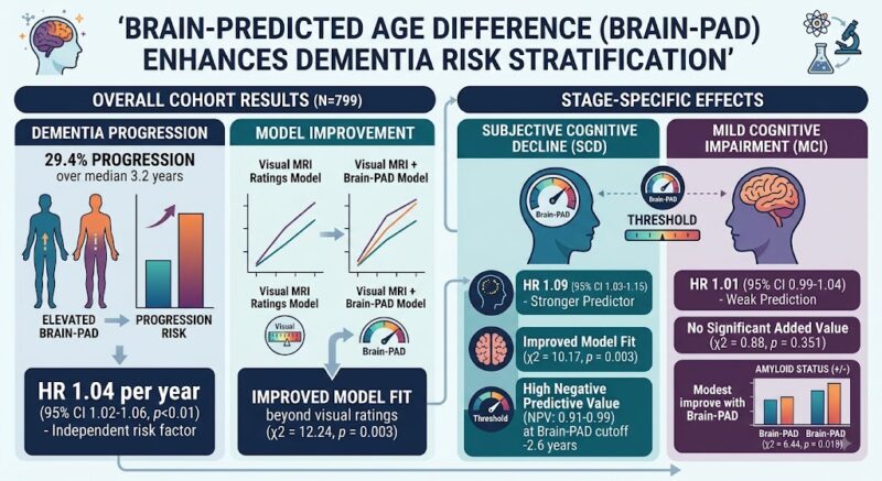
Overall, 29.4% of the cohort progressed to dementia during follow-up. Elevated brain-PAD was independently associated with increased risk of progression (hazard ratio [HR] 1.04 per year, 95% confidence interval [CI] 1.02-1.06, p<0.01) and improved model fit beyond visual MRI ratings (χ2 = 12.24, p = 0.003).
Importantly, interaction analyses revealed stage-specific effects: brain-PAD was a stronger predictor in the SCD group (HR 1.09, 95% CI 1.03-1.15) than in MCI (HR 1.01, 95% CI 0.99-1.04). In SCD patients, brain-PAD led to better model fit (χ2 = 10.17, p = 0.003) and modest gains in discrimination (C-index increased by 0.02) beyond visual MRI ratings. A threshold brain-PAD cutoff of −2.6 years identified individuals with a high negative predictive value (0.91–0.99) for dementia progression over 2 to 10 years, potentially useful for ruling out imminent risk.
In the biomarker subset, brain-PAD improved model fit beyond amyloid status alone (χ2 = 6.44, p = 0.018) but did not enhance discrimination metrics. In contrast, among MCI patients, brain-PAD offered no significant incremental predictive value (χ2 = 0.88, p = 0.351).
Expert Commentary
This study substantiates the utility of brain-PAD as a sensitive, quantitative MRI biomarker to stratify dementia risk, particularly during the early subjective phase of cognitive decline. The stronger predictive capacity in SCD suggests brain-PAD captures subtle neurodegenerative changes preceding measurable impairment. The modest improvement over amyloid biomarkers highlights brain-PAD’s complementary role alongside established molecular markers.
These findings align with biological plausibility; accelerated structural brain aging may reflect early neurodegenerative processes not fully captured by focal pathology or amyloid load. However, the lack of added value in MCI may reflect the transition to more heterogeneous pathology, where brain-PAD changes are overshadowed by other clinical and biomarker predictors.
Limitations include retrospective design and potential selection bias linked to memory clinic cohorts. Generalizability to broader populations requires prospective validation. Future integration of multimodal imaging and molecular markers may refine predictive algorithms further.
Conclusion
Brain-predicted age difference derived from structural MRI enhances early dementia risk stratification in patients with subjective cognitive decline beyond conventional visual MRI ratings and provides additional prognostic information even when amyloid status is known. Its application in clinical practice could facilitate personalized surveillance and timely therapeutic interventions. While less impactful in established mild cognitive impairment, brain-PAD represents a promising biomarker for identifying high-risk individuals at the preclinical disease stage, thus advancing precision medicine approaches in neurodegeneration.
Funding and Clinical Trial Registration
The study was supported by the Amsterdam Dementia Cohort and SCIENCe project resources. No specific clinical trial registration was reported.
References
1. De Vries S, Farkas K, Rhodius-Meester HFM, et al. Comparison of Brain Age With Standard MRI Assessment for Dementia Risk Stratification in Subjective and Mild Cognitive Impairment. Neurology. 2026 Aug 10;107(5):e218418. doi:10.1212/WNL.0000000000008418. PMID: 42574695.
2. Cole JH, Franke K. Predicting Age Using Neuroimaging: Innovative Brain Ageing Biomarkers. Trends Neurosci. 2017 Dec;40(12):681-690.
3. Jessen F, Amariglio RE, Buckley RF, et al. The characterisation of subjective cognitive decline. Lancet Neurol. 2020 Jan;19(3):271-278.
4. Petersen RC, Roberts RO, Knopman DS, et al. Mild cognitive impairment: ten years later. Arch Neurol. 2009 Dec;66(12):1447-55.
This article was created using several editorial tools, including AI, as part of the process. Human editors reviewed this content before publication.