Temporal Dynamics of Posterior Segment Imaging in Ocular Syphilis: Clinical Correlates and Laboratory Insights
Highlight
- Posterior segment imaging abnormalities in ocular syphilis vary predictably with symptom duration.
- OCT disc edema predominates in early presentations (≤8 weeks), while some lesions persist throughout disease course.
- Rapid plasma reagin titers inversely correlate with symptom duration, supporting laboratory monitoring alongside imaging.
- Temporal clustering and principal component analyses reveal distinct imaging phenotype patterns useful for clinical assessment.
Study Background
Ocular syphilis is a potentially sight-threatening manifestation of systemic Treponema pallidum infection, affecting various ocular structures, often involving the posterior segment. Due to its varied clinical presentations and resemblance to other uveitic and retinal conditions, diagnosis can be challenging. Early and accurate identification of ocular syphilis is critical to initiate appropriate antimicrobial therapy and prevent irreversible visual loss. Posterior segment imaging modalities—such as color fundus photography (CFP), fluorescein angiography (FA), optical coherence tomography (OCT), and near-infrared reflectance (NIR)—provide detailed structural and vascular information but lack standardized temporal characterization relative to symptom onset. This study aims to delineate reproducible temporal patterns of posterior segment imaging findings in ocular syphilis to enhance diagnostic precision and improve clinical management strategies.
Study Design
This retrospective case series examined patients with a confirmed diagnosis of ocular syphilis from 2015 to 2025. Inclusion criteria required documented symptom duration and availability of posterior segment imaging data. Imaging modalities analyzed consisted of CFP, FA, OCT, and NIR across 73 eyes from 40 patients, with complete multimodal imaging in 46 eyes. Posterior segment findings observed in five or more eyes were systematically graded and evaluated across discrete symptom-duration intervals. Generalized estimating equation (GEE) logistic regression accounted for intra-patient correlation when comparing imaging features between early (≤8 weeks) and later symptom presentations. Hierarchical clustering and principal component analysis (PCA) elucidated temporal patterns among imaging abnormalities. Laboratory correlates, including serum rapid plasma reagin (RPR) titers and cerebrospinal fluid (CSF) markers, were analyzed using Spearman rank correlation to investigate their relationship with symptom duration.
Key Findings
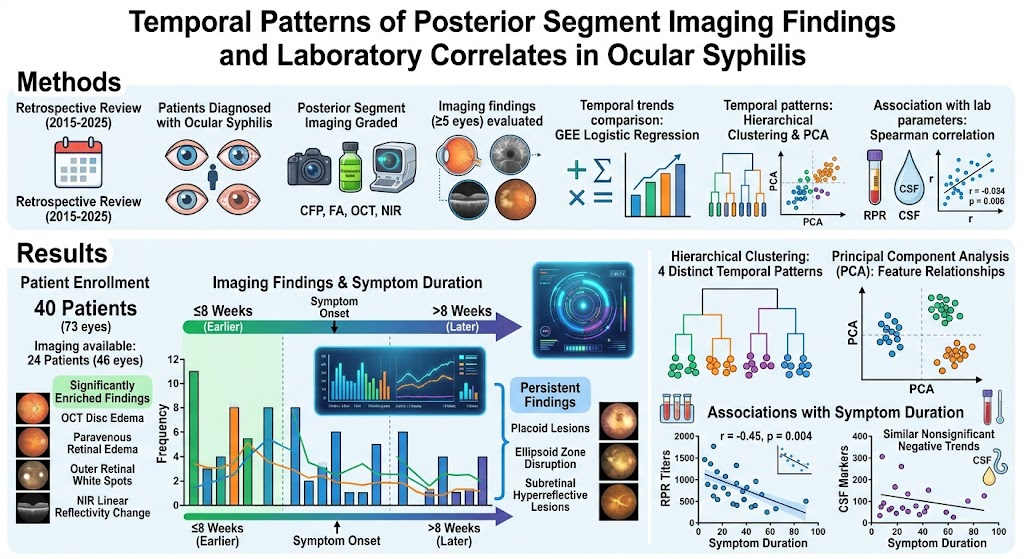
The study identified that posterior segment abnormalities were more frequently detected in patients presenting earlier after symptom onset (≤8 weeks). OCT imaging revealed disc edema significantly enriched in earlier presentations, with statistical confirmation (p-value not specified in abstract), suggesting active inflammation or optic nerve involvement predominates early in disease. Paravenous retinal edema, outer retinal white spots, and near-infrared linear reflectivity changes shared similar temporal enrichment early in the disease course, indicating these features may represent acute inflammatory or infectious retinal responses.
Conversely, other structural abnormalities, including placoid lesions typical of syphilitic chorioretinitis, ellipsoid zone disruption (affecting photoreceptor integrity), and subretinal hyperreflective lesions, were consistently observed across all symptom-duration intervals. This persistence suggests these lesions may represent chronic or sequelae changes, less influenced by timing.
Hierarchical clustering analysis of imaging features delineated four distinct temporal patterns, effectively categorizing imaging phenotypes into early-predominant, persistent, and possibly late features. Principal component analysis provided a complementary visualization framework, enhancing understanding of inter-feature relationships and temporal segregation.
Serologically, RPR titers demonstrated a moderate inverse correlation with symptom duration (Spearman r = -0.45, p = 0.004), implying higher titers are associated with earlier disease stages. CSF parameters (details not specified) showed nonsignificant negative trends with symptom duration, potentially reflecting central nervous system involvement dynamics but requiring larger studies for clarification.
Expert Commentary
This investigation adds critical insight into the natural history of posterior segment involvement in ocular syphilis, supporting the concept that certain imaging phenotypes can act as biomarkers of disease timing. The enrichment of disc edema and retinal edema findings in early phases aligns with the known inflammatory pathology during active infection. Persistent lesions such as placoid chorioretinitis and ellipsoid zone damage likely reflect ongoing structural damage despite symptom resolution.
The moderate inverse correlation between RPR titers and symptom duration reinforces current clinical understanding and highlights the value of integrating imaging and serologic data for comprehensive disease assessment. Although CSF correlation was not statistically significant, its trend warrants exploration given the overlap with neurosyphilis diagnostics.
Limitations inherent to retrospective design include potential selection bias, variable imaging follow-up intervals, and incomplete CSF data. Additionally, the modest sample size limits generalizability and stratified subgroup analyses. Prospective studies are necessary to confirm temporal imaging biomarkers and investigate their prognostic and therapeutic implications.
Conclusion
Posterior segment imaging in ocular syphilis demonstrates distinct, nonrandom temporal patterns related to symptom duration. Recognizing early-predominant features such as OCT disc edema and retinal edema can aid clinicians in diagnosing active disease phases, whereas persistent findings reflect chronic involvement. Integrating multimodal imaging findings with laboratory markers like RPR titers enhances clinical interpretation and management decisions. Future research should focus on validating these temporal phenotypes prospectively and exploring their role in monitoring treatment response and predicting visual outcomes.
Funding and Clinical Trials
The original study funding and clinical trial registration details were not provided in the source abstract.
References
1. Rhead JA, Chen A, Seddon I, Browning DJ. Temporal Patterns of Posterior Segment Imaging Findings and Laboratory Correlates in Ocular Syphilis. Am J Ophthalmol. 2026 Aug 13. PMID: 42595286.
2. Mattei PA, Pepple KL, Gonzales JA. Ocular syphilis: clinical features and management. Curr Opin Ophthalmol. 2017;28(6):607-613.
3. Koizumi H, Tateno Y, Inoue T, et al. Multimodal Imaging of Syphilitic Posterior Placoid Chorioretinitis. Ophthalmology. 2014;121(6):124-134.
4. Tang S-M, Wang J, Ichhpujani P. Optical coherence tomography in infectious uveitis: patterns and applications. Asia Pac J Ophthalmol (Phila). 2020;9(4):283-297.
5. Workowski KA, Bachmann LH, Chan PA, et al. Sexually transmitted infections treatment guidelines, 2021. MMWR Recomm Rep. 2021;70(4):1-187.
This article was created using several editorial tools, including AI, as part of the process. Human editors reviewed this content before publication.