Enhancing Obstetric Decision-Making: The Role of Intrapartum Sonography in Managing Difficult Labor
Introduction
Difficult labor, characterized by protracted or arrested cervical dilation in the first stage and arrest of fetal descent in the second, remains a significant contributor to intrapartum cesarean deliveries and instrumental birth-related maternal and neonatal morbidity. Traditional assessment relies heavily on digital vaginal examination to determine fetal head position, attitude, and station; however, this method is uncomfortable for patients, requires repetitive examinations, and suffers from substantial imprecision in critical clinical scenarios. This article explores the clinical utility of intrapartum sonography as a complementary tool in managing difficult labor, focusing on its diagnostic advantages, clinical applications, and implications for obstetric practice.
Background and Clinical Context
Digital vaginal examination in labor is the cornerstone for assessing fetal position and progress, yet it presents several limitations. Factors such as caput succedaneum (scalp swelling), fetal head molding, deflexion, and a high presenting part can obscure the palpable sutures and fontanelles, increasing subjectivity. Furthermore, the ischial spines, key landmarks for assessing fetal station, are felt rather than visualized, compounding inaccuracy. Reported discordance between digital and sonographic assessment of fetal occiput position ranges from 20% to 33%, and digital estimation of station similarly lacks precision especially in cases with substantial caput or molding.
This diagnostic uncertainty has tangible clinical consequences, influencing decisions regarding labor augmentation, timing and mode of delivery, and instrument-assisted birth. Accurate, objective data is therefore critical, particularly in difficult labor scenarios that pose heightened risks.
Intrapartum Sonography: Techniques and Measurements
Intrapartum sonography employs a curvilinear transducer, commonly available on labor wards. It involves a limited number of targeted images and is better tolerated by women compared to repeated digital examinations.
Key sonographic assessments include:
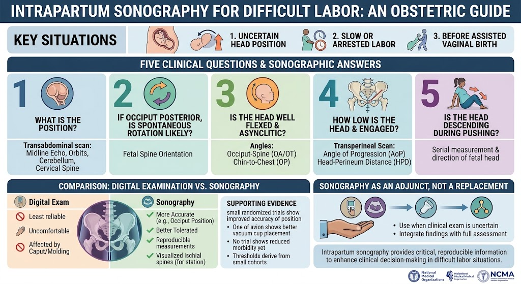
1. Fetal Head Position: Transabdominal ultrasound identifies midline cerebral echo structures such as the cavum septi pellucidi, orbits, cerebellum, and cervical spine to determine occiput position accurately.
2. Likelihood of Spontaneous Rotation: When occiput posterior is detected, fetal spine orientation suggests the probability of spontaneous rotation to an anterior position.
3. Head Attitude and Flexion: The occiput-spine angle quantifies flexion and asynclitism in occiput anterior and transverse presentations, while the chin-to-chest angle assesses flexion in occiput posterior positions.
4. Station and Engagement: Transperineal ultrasound measures the angle of progression and head-perineum distance to objectively quantify descent and engagement.
5. Descent During Pushing: Serial measurements track fetal head movement, supplemented by the directional assessment of the fetal head.
Evidence and Clinical Findings
Studies have consistently demonstrated that sonographic determination of occiput position surpasses the accuracy of digital examination. Two randomized controlled trials (RCTs) indicate that adding ultrasound enhances correct identification of fetal position prior to assisted vaginal delivery. Notably, one RCT reported improved vacuum cup placement accuracy when sonographic guidance was used.
Measurements of station and head attitude exhibit reproducibility and correlate with delivery modes, though published thresholds are often derived from modest patient cohorts and anchored against digital station assessments. Confounding factors such as transducer pressure, caput, molding, parity, and occiput position affect measurement precision.
Despite these advances, no controlled trials have definitively demonstrated that interventions guided solely by intrapartum sonographic data reduce maternal or neonatal morbidity. Therefore, sonography currently serves as an adjunct rather than a replacement for clinical examination.
Application Scenarios in Clinical Practice
The authors propose three main clinical scenarios where intrapartum sonography can influence management:
– Uncertain Fetal Head Position: When fetal head position is unclear on digital examination, ultrasound can provide definitive confirmation.
– Slow or Arrested Labor: Sonographic evaluation helps characterize head attitude, degree of flexion, and descent objectively, assisting in diagnosing causes of labor dystocia.
– Immediately Before Assisted Vaginal Birth: Sonography offers critical information to optimize instrument placement and anticipate potential difficulties.
Expert Commentary and Limitations
Integrating intrapartum sonography into routine labor management demands training, equipment availability, and standardized protocols. Although the evidence favors better diagnostic accuracy, the absence of large-scale outcome-based trials limits changes in clinical guidelines. The technique is user-dependent and factors such as fetal position complexity and operator experience can influence results.
Furthermore, variations in sonographic thresholds highlight the need for larger, multicenter studies to establish standardized measurement criteria with proven clinical benefit.
Conclusion and Future Directions
Intrapartum sonography represents a valuable adjunct in the evaluation of difficult labor by enhancing accuracy in assessing fetal head position, attitude, and station. Its implementation may improve decision-making surrounding labor management and assisted vaginal birth, thereby potentially reducing procedure-related morbidity.
Nevertheless, sonography should complement, not replace, clinical examination. Future research must focus on robust randomized trials to evaluate whether sonography-guided management improves maternal and neonatal outcomes and to define standardized sonographic criteria for labor management.
Continued education and integration of intrapartum sonographic skills for obstetricians and laborists will be critical for translating these advances into routine practice.
Reference
Mappa I, Rizzo G. Intrapartum Sonography for Difficult Labor. Am J Obstet Gynecol. 2026 Aug 14:S0002-9378(26)00415-1. doi: 10.1016/j.ajog.2026.08.012. Epub ahead of print. PMID: 42600675.
This article was created using several editorial tools, including AI, as part of the process. Human editors reviewed this content before publication.